Ex vivo analysis of renal proximal tubular cells
Résumé
Background : Experimental models are inevitably a compromise between accurately reproducing a pathological situation and schematically simplifying it, which is intended to provide both relevance and conclusiveness. In-vivo models are very relevant, but multiple cell-types undergoing various changes may hinder the observation of individual molecular events.
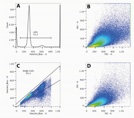
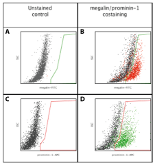
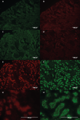
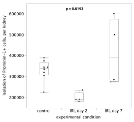
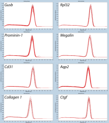
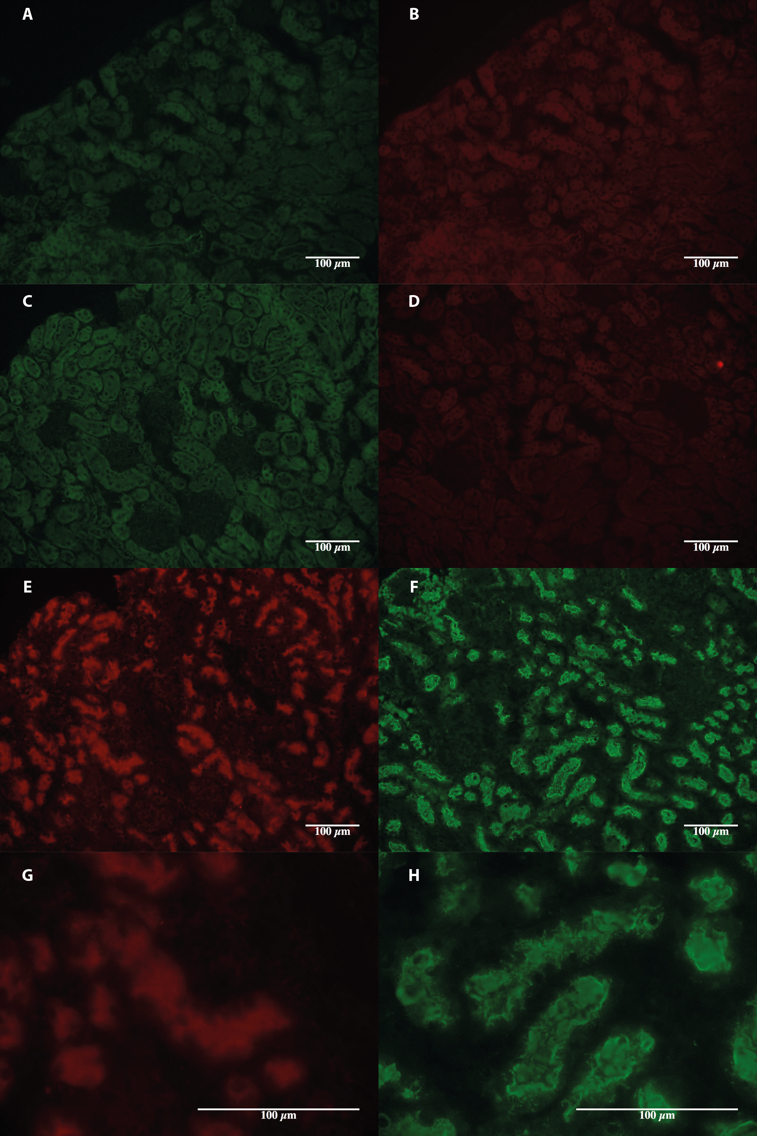
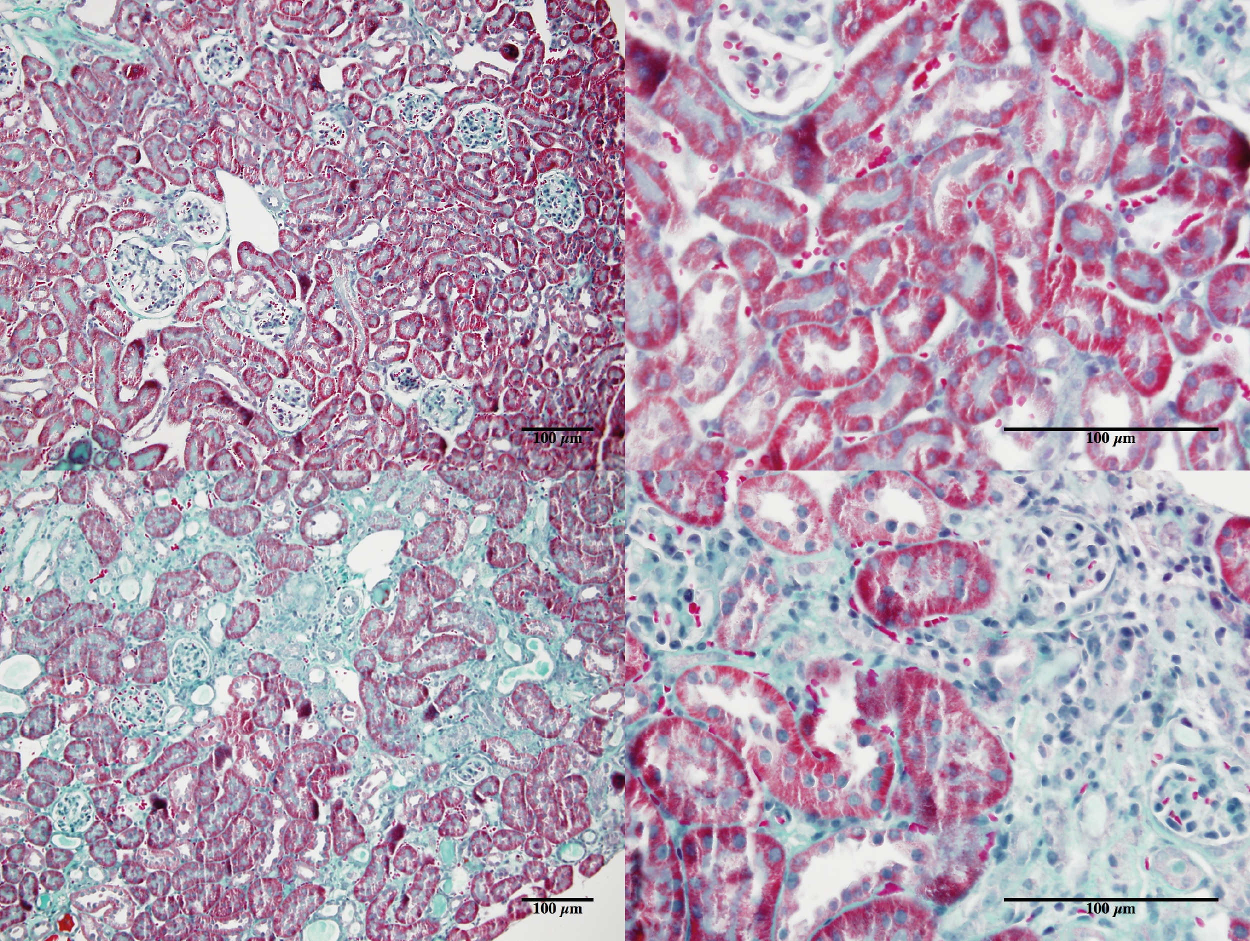
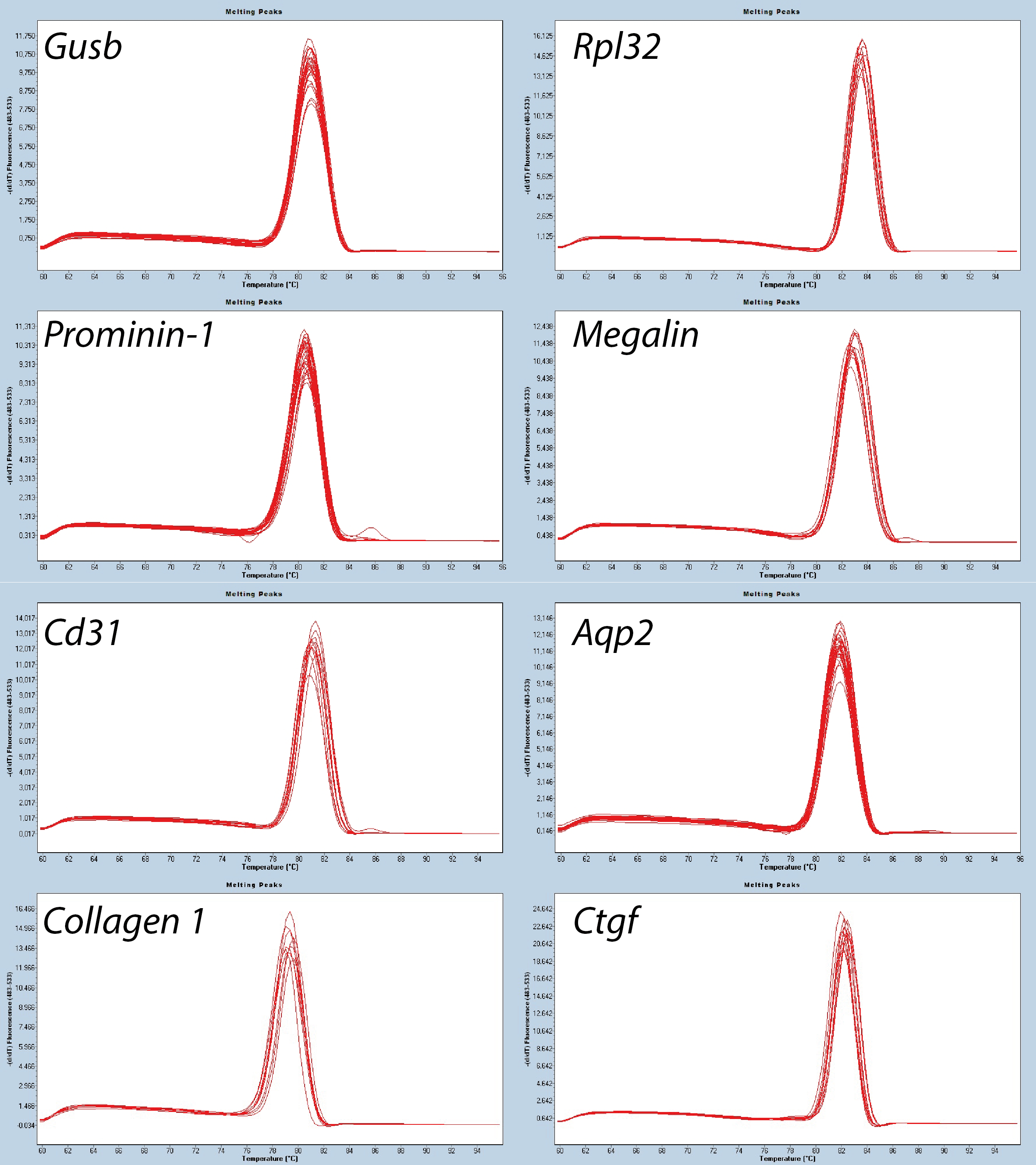
Results : Here, we describe a method for analyzing and isolating specific cell types from the kidney and studying the phenotype they have acquired in vivo. Using flow cytometry, immunofluorescence, and RT-PCR, we show that our method is suitable for studying and isolating proximal tubular cells with an anti Prominin-1 antibody. Kidneys are subjected to mechanical dissociation followed by flow-cytometry analysis. Hundreds of thousands of proximal tubular cells are then isolated by magnetic separation followed by direct analysis or primary cell culture. Using our method, we detect phenotypic changes in the proximal tubular cells after renal ischemia reperfusion, and we isolate the proximal tubular cells, with a purity over 80%.
Conclusions : This method is efficient, quick, simple, and cheap, and should be useful for studying cell-type specific parameters after in vivo experimental studies. It is also a simple method to obtain a specific primary cell culture from any animal strain.
Fichier principal
 Legouis_2015_Ex_vivo_analysis_of.pdf (2 Mo)
Télécharger le fichier
Legouis_2015_Ex_vivo_analysis_of.pdf (2 Mo)
Télécharger le fichier
 s12860-015-0058-4-s1.png (3.54 Mo)
Télécharger le fichier
s12860-015-0058-4-s1.png (3.54 Mo)
Télécharger le fichier
 s12860-015-0058-4-s2.png (713.88 Ko)
Télécharger le fichier
s12860-015-0058-4-s2.png (713.88 Ko)
Télécharger le fichier
 s12860-015-0058-4-s3.png (10.88 Mo)
Télécharger le fichier
s12860-015-0058-4-s3.png (10.88 Mo)
Télécharger le fichier
 s12860-015-0058-4-s4.png (8.14 Mo)
Télécharger le fichier
s12860-015-0058-4-s4.png (8.14 Mo)
Télécharger le fichier
 s12860-015-0058-4-s5.png (160.23 Ko)
Télécharger le fichier
s12860-015-0058-4-s6.docx (14.81 Ko)
Télécharger le fichier
s12860-015-0058-4-s5.png (160.23 Ko)
Télécharger le fichier
s12860-015-0058-4-s6.docx (14.81 Ko)
Télécharger le fichier
 s12860-015-0058-4-s7.png (747.76 Ko)
Télécharger le fichier
s12860-015-0058-4-s7.png (747.76 Ko)
Télécharger le fichier
Origine : Publication financée par une institution
Format : Figure, Image
Origine : Fichiers produits par l'(les) auteur(s)
Origine : Fichiers produits par l'(les) auteur(s)
Format : Figure, Image
Origine : Fichiers produits par l'(les) auteur(s)
Origine : Fichiers produits par l'(les) auteur(s)
Format : Figure, Image
Origine : Fichiers produits par l'(les) auteur(s)
Origine : Fichiers produits par l'(les) auteur(s)
Format : Figure, Image
Origine : Fichiers produits par l'(les) auteur(s)
Origine : Fichiers produits par l'(les) auteur(s)
Format : Figure, Image
Origine : Fichiers produits par l'(les) auteur(s)
Origine : Fichiers produits par l'(les) auteur(s)
Origine : Fichiers produits par l'(les) auteur(s)
Format : Figure, Image
Origine : Fichiers produits par l'(les) auteur(s)
Origine : Fichiers produits par l'(les) auteur(s)
Loading...

