Ex vivo porcine model for eye, eyelid, and orbit movement analysis of 4-mm ferromagnetic foreign bodies in MRI
Résumé
Purpose Ferromagnetic foreign bodies (FFB) present during magnetic resonance imaging (MRI) explorations can lead to tissue injury due to movement, especially in and around the eyes. Ferromagnetic foreign bodies located in the intraocular area, eyelids, and orbit are thus prohibited from undergoing MRI. The aim of the study was to analyze movement of 4-mm ferromagnetic foreign bodies in MRI in the eye, eyelid, and orbit using computed tomography (CT) scan. Method We developed a porcine model using 12 quarters of fresh porcine heads. Each porcine head included one whole orbit with the ocular globe, orbital fat, muscles, and eyelids. Four-millimeter FFB were implanted in the eye within 2 days post-slaughter, and images were acquired within 5 days post-slaughter. Four-millimeter FFB movement was analyzed after 1.5-Tesla (T) MRI. Four locations were tested: intravitreous, suprachoroidal, intraorbital fat, and intrapalpebral. Movement analysis was assessed using computed tomography (CT) scan. Results The intravitreous ferromagnetic ball moved 14.0 +/- 8.8 mm (p < 0.01), the suprachoroidal ball moved 16.8 +/- 5.4 mm (p < 0.01), the intraorbital fat ball moved 5.8 +/- 0.9 mm (p > 0.05), and the intrapalpebral ball moved 2.0 +/- 0.4 mm (p > 0.05). Conclusion The ex vivo porcine model was able to study FFB movement. The 4-mm ferromagnetic balls moved in intravitreous and in suprachoroidal locations after MRI.
Domaines
Sciences du Vivant [q-bio]
Fichier principal
 Ghemame et al - 2021 - Ex vivo porcine model for eye, eyelid -Revised manuscript clean copy.pdf (988 Ko)
Télécharger le fichier
Ghemame et al - 2021 - Ex vivo porcine model for eye, eyelid -Revised manuscript clean copy.pdf (988 Ko)
Télécharger le fichier
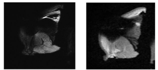 Fig Annexe.jpg (43.81 Ko)
Télécharger le fichier
Fig Annexe.jpg (43.81 Ko)
Télécharger le fichier
Origine : Fichiers produits par l'(les) auteur(s)
Format : Figure, Image