Enhancing Eye Fundus Images for Diabetic Retinopathy Screening
Résumé
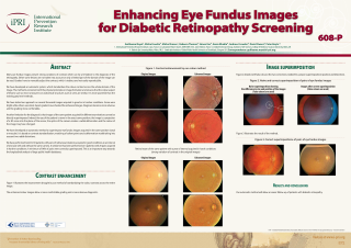
Many eye fundus images present strong variations of contrast which can be a limitation to the diagnosis of the retinopathy. Either some lesions are not taken into account or only a limited part of the domain of the image can be read. Graders have to manually adjust the contrast, which is tedious and not easily reproducible. We have developed an automatic system, which standardises the colour contrast across the whole domain of the image. The method is consistent with the physical principles or image formation and ensures that the colour aspect of lesions such as micro-aneurysms or anatomical structures such as veins are similar. It is more powerful than the existing grey level methods. We have tested our approach on several thousand images acquired in good or in harsher conditions. Some were bright while others were dark. Expert graders have checked the enhanced images. Diagnosis becomes more obvious and the grading more comfortable. Another limitation for the diagnosis is that images of the same patient acquired for different examinations cannot be directly superimposed. Indeed, the eye of the patient is never in the exact same position, the image is a projection of a 3D scene into the plane of the sensor, the optics of the camera creates a radial deformation and the colour of the image may have changed. We have developed an automatic method to superimpose eye fundus images acquired in the same position (nasal or macular). It is based on contrast standardisation, matching of salient points and a deformation model taking into account two radial distortions. We have performed tests for 69 patients with pairs of retinal examinations acquired in good conditions at an interval of one year with and without the same camera. A similar test has been performed on 5 patients with 20 pairs acquired in harsher conditions. A minimum of 96% of pairs were correctly superimposed. This is an important step towards the longitudinal analysis of large public health databases.
Fichier principal
 2017_NoyelOwensBoyle_ADA_poster_Hal.pdf (8.65 Mo)
Télécharger le fichier
2017_NoyelOwensBoyle_ADA_abstract_src.pdf (461.15 Ko)
Télécharger le fichier
2017_NoyelOwensBoyle_ADA_poster_Hal.pdf (8.65 Mo)
Télécharger le fichier
2017_NoyelOwensBoyle_ADA_abstract_src.pdf (461.15 Ko)
Télécharger le fichier
Origine : Fichiers produits par l'(les) auteur(s)
Origine : Fichiers produits par l'(les) auteur(s)