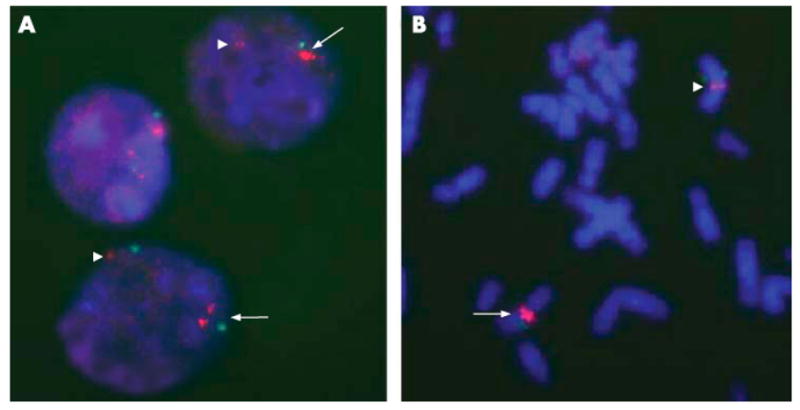
Figure 2.
FISH analysis of the duplication, showing three specific 7q11-probe sites on interphase cells (A) and two hybridization spots on metaphase chromosomes (B). The normal signal is indicated by arrowheads and the duplication by arrows. Magnification x 1000.