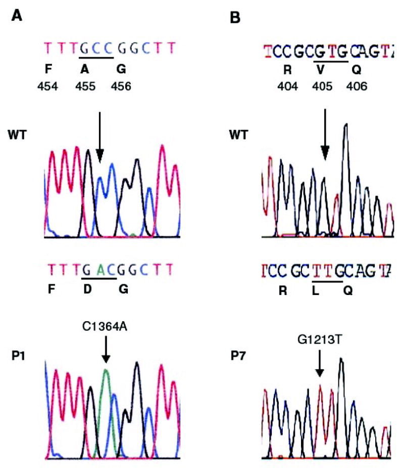
Figure 3.
Sequence chromatograms from normal individual (wild type, WT) and affected individuals (P) are shown together with the expected amino-acid changes. Nucleotide variations are indicated by an arrow. A: 1364C>A inducing the A455D mutation in Patient 1 (P1), B: 1213G>T inducing the V405L mutation in Patient 7 (P7).